Published in Frontiers in Human Neuroscience in 2022, this review intended to answer how ultrasound for neuromodulation works
Our review titled, Current state of potential mechanisms supporting low intensity focused ultrasound for neuromodulation, attempts to address the following questions: 1) How can we alter the amount of mechanical energy or other properties of the mechanical energy using the sonication parameters available with each device, 2) How are neuronal tissue affected by mechanical energy, and 3) How do those sonication parameters change the type of neuromodulation (i.e., excitatory or suppressive)? We reviewed the theoretical mechanisms of action for neuromodulation and the empirical findings tracking all the sonication parameters used to elucidate the possible link between the proposed mechanisms of action and the choice of sonication parameters. This is still an emerging field, but a tabulation of the empirical findings and theoretical models is needed to help clinicians and researchers choose the best paradigm to use.
Get The Article
- Article
- Readcube
Frontiers in Human Neuroscience: Brain Imaging and Stimulation Open Source Article
Current State of Potential Mechanisms Supporting Low-Intensity Focused Ultrasound for Neuromodulation PDF
Cite This Work
- MLA
- APA
- Chicago
- Harvard
- Vancouver
DellItalia, John, et al. “Current state of potential mechanisms supporting low intensity focused ultrasound for neuromodulation.” Frontiers in Human Neuroscience: 228.
DellItalia, J., Sanguinetti, J. L., Monti, M. M., Bystritsky, A., & Reggente, N. Current state of potential mechanisms supporting low intensity focused ultrasound for neuromodulation. Frontiers in Human Neuroscience, 228.
DellItalia, John, Joseph L. Sanguinetti, Martin M. Monti, Alexander Bystritsky, and Nicco Reggente. “Current state of potential mechanisms supporting low intensity focused ultrasound for neuromodulation.” Frontiers in Human Neuroscience: 228
DellItalia, J., Sanguinetti, J.L., Monti, M.M., Bystritsky, A. and Reggente, N., Current state of potential mechanisms supporting low intensity focused ultrasound for neuromodulation. Frontiers in Human Neuroscience, p.228.
DellItalia J, Sanguinetti JL, Monti MM, Bystritsky A, Reggente N. Current state of potential mechanisms supporting low intensity focused ultrasound for neuromodulation. Frontiers in Human Neuroscience.:228.
Science Without Jargon
Science should be accessible to everyone. However, dense jargon-filled articles can make it difficult for non-experts to engage with research. Making science accessible promotes scientific literacy and informed decision-making. In this post, we summarize our recent article for a lay audience.
How ultrasound for neuromodulation works
An alternative to the putative non-invasive brain stimulation is devices using ultrasound. Ultrasound has been used for decades by clinicians to image various parts of the body, but recently ultrasound devices have begun to be used for neuromodulation. Ultrasound doesn’t use electric or magnetic fields, rather it generates acoustic waves that are a mechanical force. This mechanical force can be focused on a precise area with only the maximal mechanical energy converging on millimeter-sized region. This allows for deeper and/or smaller brain regions to be targeted compared to transcranial electrical stimulation and transcranial magnetic stimulation. However, the different energy source compared to electric or magnetic fields requires a different understanding of how neuromodulation occurs. Without this understanding, effective uses of ultrasound in empirical research and clinical practices will be limited.
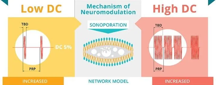
Ultrasound’s acoustic waves have the characteristic properties of wavelength, amplitude, and frequency. Wavelength is the distance between two peaks within the wave, the amplitude is the height of the wave, and frequency is the number of peaks in a second. Each of these properties affects the total amount of mechanical energy delivered by the ultrasound device and other sonication parameters. The total energy can be measured by either the average amount in a spatial region or the average amount delivered over time typically converted into the units of watts per centimeter squared. In addition to the intensity, the total energy delivered over time is affected by the duty cycle, which is the percentage of time the sonication occurs. The duty cycle also determines if a paradigm is pulsed or continuous. Pulsed paradigms are any duty cycle below 100 percent, which allows for breaks between the sonication, compared to a continuous application of ultrasound. The frequency of the ultrasound’s acoustic waves is related to the sonication parameters of center frequency and pulse repetition frequency. The center frequency is set by the device manufacturer, which is the frequency delivered by the device and this frequency is related to the spatial precision of the acoustic wave delivered. The pulse repetition frequency is the frequency of the acoustic wave delivered by the pulsed paradigm. The final commonly adjusted sonication parameter is sonication duration (i.e., total time of acoustic wave delivered).
The mechanical energy delivered by the ultrasound device has seven proposed mechanisms to affect the activity of groups of neurons. Neurons are connected and each neuron’s activity either helps to excite other neurons connected to it or suppresses the activity of the neurons connected to it. These signals involve both electrical and chemical signaling. Since ultrasound is mechanical, the mechanisms of action proposed describes: 1) effects of mechanical energy on the temperature., 2) how the neurons detect and transform that mechanical energy to electrical or chemical signaling (i.e., mechanosensitive ion channels), or 3) how mechanical energy interacts with the elasticity of neurons to change the electrical properties or structure of neurons (e.g., direct flexoelectricity, change of membrane conformational states, or sonoporation).
Ultrasound has been used for decades to destroy malignant tissue by using enough intensity to generate larger changes in temperature. These large changes in temperature are not seen in the intensity ranges used in non-invasive brain stimulation. Despite the lower intensity used, there is still a mechanical force acting on the neurons. Some neurons have specific mechanisms for detecting external mechanical forces. These are most well understood in our tactile sensations. When our hand presses against a surface, specialized neurons detect the mechanical force by getting stretched which allows for chemical and electrical signaling to occur. The amount and distribution of neurons with similar properties in the brain is an active area of research. In addition to these specialized neurons, the mechanical energy from ultrasound can change the electrical properties of neurons by distorting the shape. The specific configuration of the membrane allows for electrical signals (i.e., direct flexoelectricity) to be produced as the mechanical energy changes the alignment of the interior and exterior parts of the membrane. On top of these alignment changes, there are pressure changes which can also generate both chemical and electrical changes from the changes in membrane conformational states.
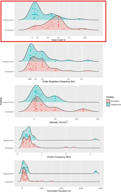
Additionally, the neuron’s membranes can have changes to their permeability called sonoporation allowing for electrical changes that can elicit the neuron to fire. This process was investigated how the ultrasound’s pulse repetition frequency, intensity, or duty cycle could produce excitatory or suppressive effects. The key sonication parameter that best predicted differences in neuronal activity was duty cycle. Higher duty cycles between 10% to 70%, typically excited neurons, while lower duty below 10% created suppressive effects. Unfortunately, this one parameter did not predict the suppressive findings well, but the excitatory findings were almost all exclusively found between 10% to 70% duty cycle.
While duty cycle was predictive of some results found in the literature, it left most of the results unexplained. More models and theories are needed to expand the understanding of the mechanisms of action. Hopefully, this review gives a basic knowledge base to clinicians and researchers to use in their treatments or experiments. As the understanding of the mechanisms of action expand, more nuanced treatments and experiments can be used.
Figures & Captions
Feel free to use these figures in your articles, blogs, and presentations. If you do, please cite this work.
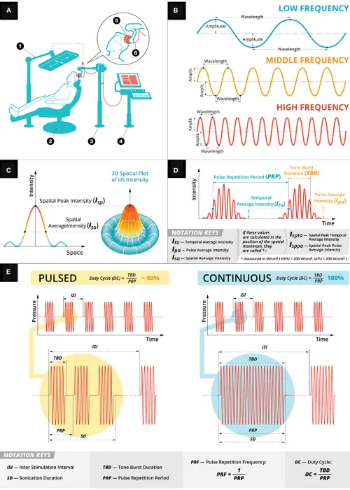
Figure 1.

Figure 2.

Figure 3.