VR for Cognition and Memory
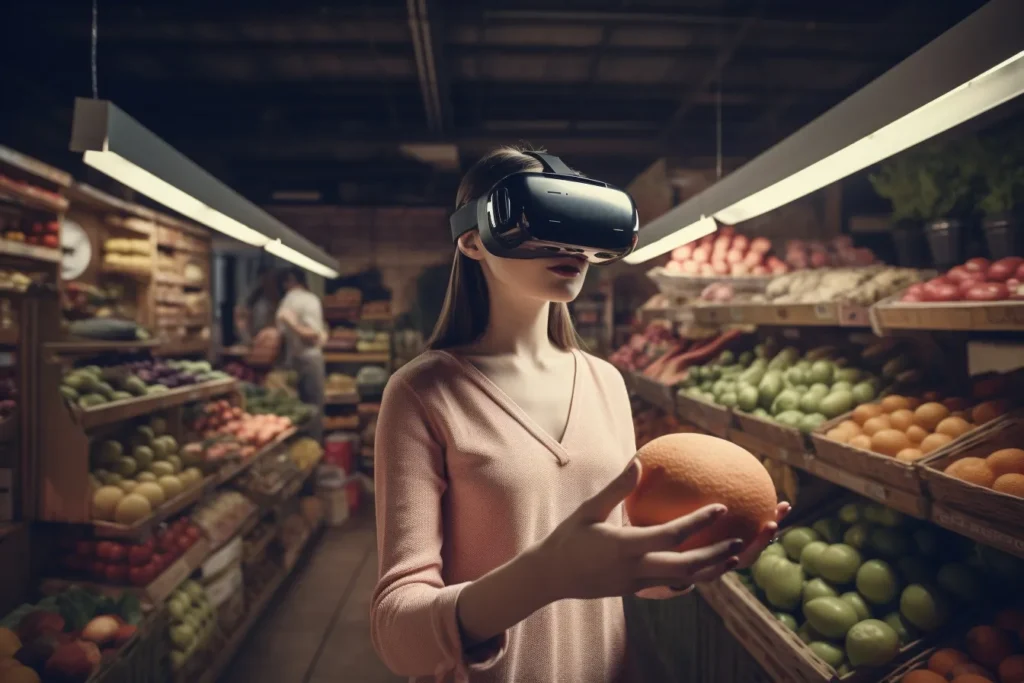
Get The Article PDF Paywalled Chapter VR for Cognition and Memory Cite This Work APA AMA MLA Reggente N. (2023). VR for Cognition and Memory. Current topics in behavioral neurosciences, 10.1007/7854_2023_425. Advance online publication. https://doi.org/10.1007/7854_2023_425 Reggente N. VR for Cognition and Memory [published online ahead of print, 2023 Jul 14]. Curr Top Behav Neurosci. 2023;10.1007/7854_2023_425. doi:10.1007/7854_2023_425 Reggente, […]
Individual Differences in Aesthetic Chills
Get The Article Article PDF Scientific Data Article ChillsDB 2.0: Individual Differences in Aesthetic Chills Among 2,900+ Southern California Participants Cite This Work APA MLA Bibtex Schoeller, F., Moore, L., Lynch, C., & Reggente, N. (2023c). ChillsDB 2.0: Individual Differences in aesthetic chills among 2,900+ Southern California participants. Scientific Data, 10(1). https://doi.org/10.1038/s41597-023-02816-6 Schoeller, Felix, et al. “ChillsDB […]
Predicting Chills – Characterizing Individual Differences in Peak Emotional Response
Get The Article Article PDF PNA Nexus Article Predicting individual differences in peak emotional response Cite This Work APA MLA Bibtex Schoeller, F., Christov-Moore, L., Lynch, C., Diot, T., & Reggente, N. (2024). Predicting Individual Differences in Peak Emotional Response. PNAS Nexus, 3(3). https://doi.org/10.1093/pnasnexus/pgae066 Schoeller, Félix, Leonardo Christov-Moore, et al. “Predicting Individual Differences in Peak Emotional Response.” PNAS […]
Current State of Potential Mechanisms Supporting Low-Intensity Focused Ultrasound for Neuromodulation
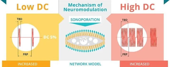
Published in Frontiers in Human Neuroscience in 2022, this review intended to answer how ultrasound for neuromodulation works Our review titled, Current state of potential mechanisms supporting low intensity focused ultrasound for neuromodulation, attempts to address the following questions: 1) How can we alter the amount of mechanical energy or other properties of the mechanical energy using the sonication […]
The Consciousness Compass
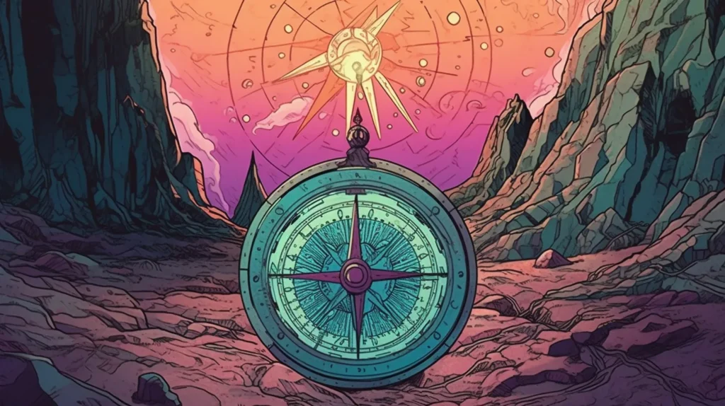
The Consciousness Compass Edited by GPT 4. Illustrated by MidJourney. What does this mean? See Afterword below. What the hell is ‘a common denominator of consciousness’”? you ask me. We’re in a café in the city on a rainy day. You’ve just opened my new paper on your laptop, recently published in the peer-reviewed Nature Portfolio journal Biology […]
Arousal Regulation by the External Globus Pallidus: A New Node for the Mesocircuit Hypothesis
Get The Article Article PDF Brain Sciences Article Arousal Regulation by the External Globus Pallidus: A New Node for the Mesocircuit Hypothesis Cite This Work APA MLA Bibtex Zheng, Z. S., Reggente, N., & Monti, M. M. (2023). Arousal regulation by the external globus pallidus: a new node for the mesocircuit hypothesis. Brain Sciences, 13(1), 146. https://doi.org/10.3390/brainsci13010146 […]
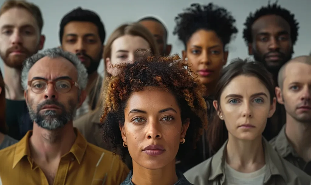
In Pursuit of a ‘Neuroprint’ for Neuroscience-Based Personalization

The Challenge of Individual Variation Just as fingerprints represent multivariate patterns with sufficient uniqueness and stability to serve as forensic evidence in criminal proceedings, our brains harbor distinctive topographies of response to the world around us. These neural landscapes, shaped by genetics, experience, and countless moments of conscious and unconscious processing, hold the key to […]
Virtual Reality vs. Reflective Chamber: New Study Shows Both Can Help Reduce Anxiety
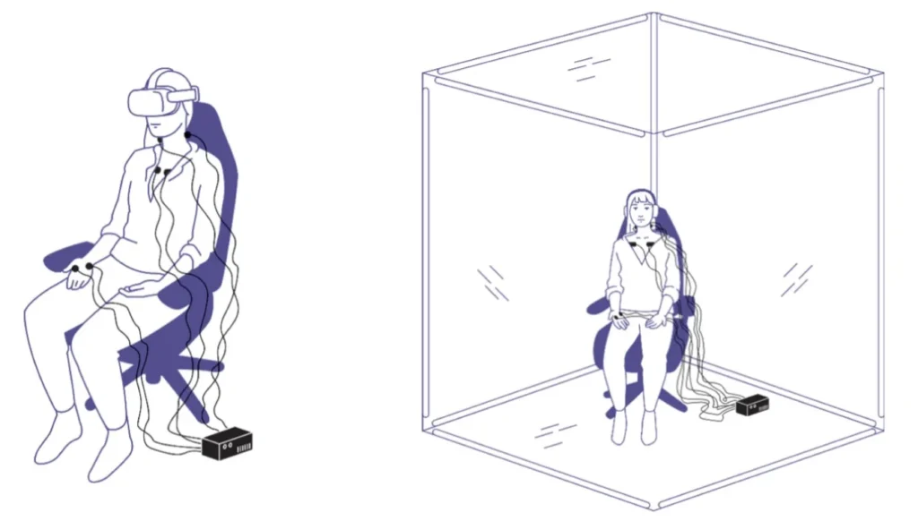
Get The Article Article PDF PLOS Mental Health Article Contrasting cognitive, behavioral, and physiological responses to breathwork vs. naturalistic stimuli in reflective chamber and VR headset environments Cite This Work APA MLA Bibtex Simonian, N., Johnson, M. A., Lynch, C., Wang, G., Kumaravel, V., Kuhn, T., Schoeller, F., & Reggente, N. (2025). Contrasting cognitive, behavioral, […]
Decoding Depth of Meditation: EEG Insights from Expert Vipassana Practitioners
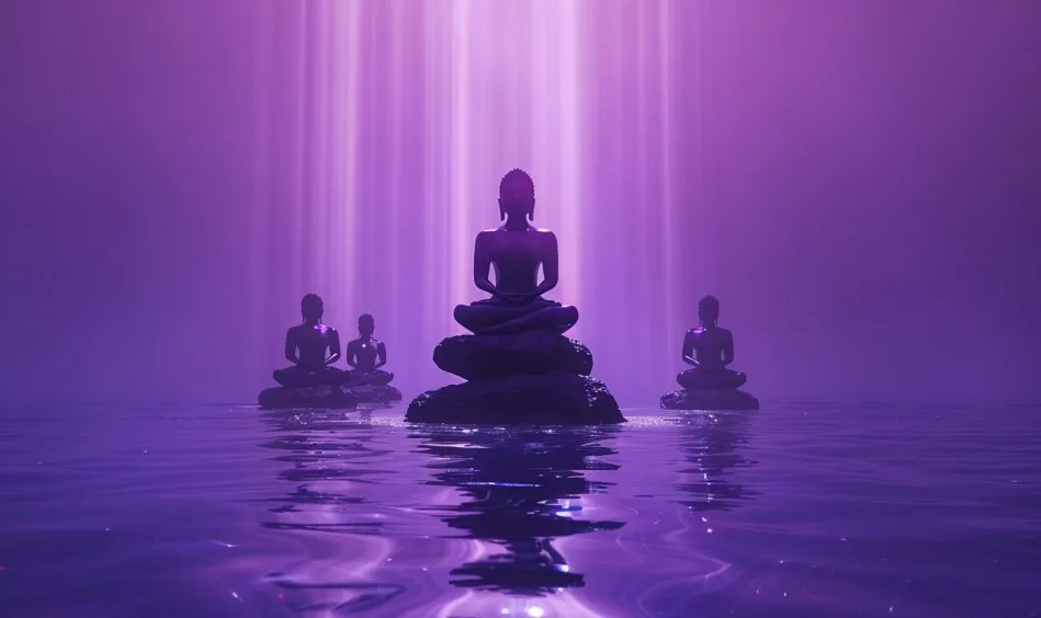
Get The Article Article PDF Biological Psychiatry Article Decoding Depth of Meditation: Electroencephalography Insights From Expert Vipassana Practitioners Cite This Work APA MLA Bibtex Reggente, N., Kothe, C., Brandmeyer, T., Hanada, G., Simonian, N., Mullen, S., & Mullen, T. (2024). Decoding Depth of Meditation: EEG Insights from Expert Vipassana Practitioners. Biological Psychiatry Global Open Science, […]